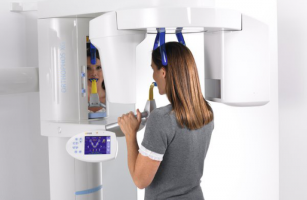
Welcome to the Future: What 3-D Dental X-rays Mean for You

What if your dentist told you that X-rays can be done in a single sweep, expose you to less radiation than a roundtrip flight from Paris to Tokyo, and result in a complete, 3-D picture of your mouth in as little as a few seconds? For anyone who has ever had a mouthful of bitewings, it’s a dream come true, thanks to a relatively new technology called “cone beam radiography”. If you’ve never heard of it, you could be missing out. Here’s the scoop on this revolutionary diagnostic tool and what it could mean for your oral health.
What Makes Cone Beam Radiography Different?
Unlike traditional X-rays, which require exposure for one flat image, cone beam radiography – also known as cone beam computed tomography, or “CBCT” – can yield hundreds of images from just a single scan. Rather than having a beam pointed to a specific area of the mouth, a machine rotates around the patient (similar to CT scanners used in the medical field), capturing panoramic images of the mouth using a cone-shaped beam.
In addition to a much simpler and productive process, other major differences include:
- Image quality: no distortion issues exist as are typical with 2D X-rays
- Format: images are captured and stored digitally, making it easy to share or duplicate
- Cost: compared to a medical CT scan, a CBCT scan can cost up to 50% less
- Level of exposure: typically more than 2D X-rays, but less than a medical CT scan
As with traditional X-rays, CBCT scans will only be recommended when necessary, and there are standard procedures in place to minimize your exposure to radiation.
Diagnostic Advantages of a CBCT Scan
The richness, clarity, and accuracy of data that cone beam radiography provides can make a world of difference to the nature and effectiveness of your dental treatment. While 2-D X-rays provide ample information for standard practices such as detecting and treating cavities, the depth and conclusiveness of CBCT-derived data can point to the optimal approach for more complex matters such as:
- Jaw disorders: a complete view from the neck to the sinuses can yield critical insights
- Orthodontic work: unlimited angles can help guide when and where to move teeth
- Implants: added precision boosts accuracy and ease of placement and restoration
- Root canals: high-resolution images help ensure thorough removal of infected pulp
- Wisdom teeth: granular details can help dentists avoid vital nerves upon extraction
In general, many dentists also find CBCT scans to be much more effective in diagnosing tooth pain than 2D X-rays. Getting the diagnosis correct the first time can save patients from unnecessary costs and health complications down the road.
Is it Right For You?
Speak with your dentist to find out whether a CBCT scan makes sense for you. Depending on your unique dental needs and the amount of documentation your dentist already has, she can advise if cone beam radiography is necessary. If CBCT scans are not available at your dentist’s office, a medical CT scan by your local hospital or clinic may be recommended.
Sources:
CBCT & endodontics: Get it right the first time with CBCT. (n.d.). Retrieved July 17, 2015, from http://www.dentaleconomics.com/articles/print/volume-101/issue-7/features/cbct-endodontics-get-it-right-the-first-time-with-cbct.html
Dental Cone-beam Computed Tomography. (2014, June 10). Retrieved on July 18, 2015, from http://www.fda.gov/Radiation-EmittingProducts/RadiationEmittingProductsandProcedures/ MedicalImaging/MedicalX-Rays/ucm315011.htm
Patient Benefits. (2006). Retrieved July 18, 2015, from http://www.conebeam.com/patient
Peck, Jerome. (n.d.). Getting the Full Picture With Cone Beam Dental Scans Retrieved July 3, 2015, from http://www.deardoctor.com/articles/getting-the-full-picture-with-cone-beam-dental-scans/page2.php